Lumbosacral Plexus

What is the Lumbosacral Plexus?
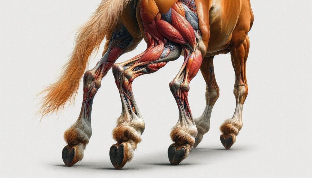
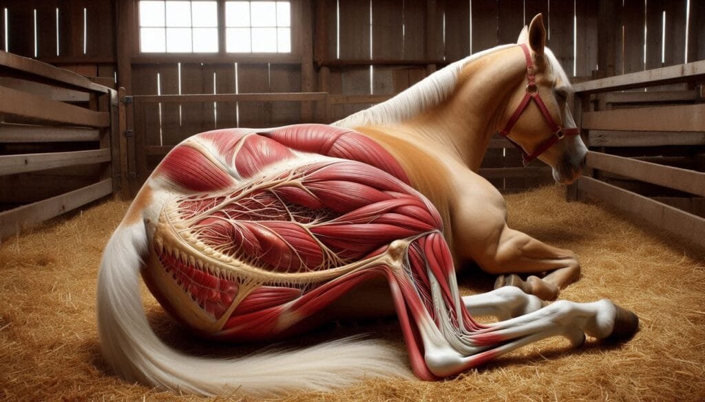
The lumbosacral plexus is formed by the ventral branches of the last three lumbar spinal nerves (L4-L6) and the first two or three sacral spinal nerves (S1-S3). This network of nerves innervates the pelvic limbs, allowing for movement and coordination.
Anatomy of the Lumbosacral Plexus
In mammals, including dogs, cats, cattle, and sheep, the lumbosacral plexus has a similar structure but varies slightly among species. Understanding this anatomy helps veterinarians diagnose and treat conditions related to nerve injuries or diseases.
Domestic Animals
In domestic mammals like dogs and cats, the lumbosacral plexus is essential for hind limb function. The major nerves originating from this plexus include:
- Ischiadic Nerve: This nerve supplies muscles involved in locomotion.
- Pudendal Nerve: It innervates pelvic organs.
- Femoral Nerve: This nerve provides motor supply to thigh muscles.
These nerves ensure coordinated movement and sensory feedback from the hind limbs. For more detailed anatomical insights, you can refer to Veterinary Anatomy.
Farm Animals
In farm animals such as cattle and sheep, the lumbosacral plexus also plays a critical role. The configuration may vary due to differences in vertebral numbers across species. However, its primary function remains consistent: to control hind limb movement.For instance, in cattle, variations in lumbar spinal nerves can influence how they walk or run. Understanding these differences is crucial for effective veterinary care. You can learn more about this from Animal Physiology.
Comparative Anatomy Across Species
The anatomy of the lumbosacral plexus can differ significantly between species. Here’s a brief comparison:
- Cats: The LSP is formed by L4-L6 and S1-S3.
- Dogs: Similar structure with variations in nerve branching.
- Cattle: May include additional contributions from thoracic nerves.
- Birds: In birds like barn owls, ten synsacral spinal nerves form their version of the plexus.
Understanding these variations helps veterinarians tailor treatments for specific species. For further reading on comparative anatomy, check out Comparative Veterinary Anatomy.
Functions of the Lumbosacral Plexus
The primary function of the lumbosacral plexus is to control muscle movements in the hind limbs. This control is essential for various activities such as walking, running, jumping, and maintaining balance.
Motor Functions
The motor functions of the lumbosacral plexus involve several key muscles:
- Quadriceps Femoris: This muscle extends the knee.
- Hamstrings: These muscles flex the knee and extend the hip.
- Gluteal Muscles: They are crucial for hip stabilization.
Damage to any part of this nerve network can lead to significant mobility issues. For more information on muscle innervation, visit Muscle Anatomy.
Sensory Functions
In addition to motor control, the lumbosacral plexus also has sensory functions. It carries sensory information from the skin and muscles back to the central nervous system. This feedback helps animals respond to their environment effectively.
Pain Perception
Nerves from this plexus play a role in pain perception as well. If an animal injures its hind limb, signals travel through these nerves to alert the brain about pain or discomfort. Understanding this process can help veterinarians manage pain effectively.
Autonomic Functions
The lumbosacral plexus also contributes to autonomic functions such as bladder control. The pudendal nerve innervates muscles that control urination. Dysfunction here can lead to serious health issues.
Clinical Significance
Understanding the lumbosacral plexus is vital for diagnosing and treating various conditions affecting animals.
Common Conditions Related to LSP Dysfunction
- Hind Limb Weakness: Weakness can result from injuries or diseases affecting any part of this nerve network.
- Loss of Coordination: Damage may lead to ataxia or uncoordinated movements.
- Urinary Incontinence: Issues with bladder control often stem from pudendal nerve dysfunction.
Veterinarians often perform neurological examinations to assess these conditions. For more details on veterinary diagnostics, see Veterinary Neurology.
Surgical Considerations
Surgeons must be aware of the lumbosacral plexus during surgeries involving the pelvis or hind limbs. Accidental damage can lead to severe complications. Techniques such as ultrasound-guided nerve blocks can help minimize risks during procedures.
Rehabilitation and Recovery
After injuries involving the lumbosacral plexus, rehabilitation plays a crucial role in recovery.
Physical Therapy
Physical therapy can help restore strength and mobility in affected animals. Techniques may include:
- Range-of-motion exercises
- Strength training
- Balance exercises
Veterinarians often recommend working with certified animal rehabilitation specialists for optimal results. You can find more information on rehabilitation at Animal Rehabilitation.
Pain Management
Effective pain management is essential during recovery. Veterinarians may prescribe medications or recommend alternative therapies like acupuncture or laser therapy.
Conclusion
The lumbosacral plexus is a critical component of animal anatomy that influences movement and overall health. Understanding its structure and function helps veterinarians provide better care for both domestic pets and farm animals. By recognizing signs of dysfunction early on, pet owners can ensure their animals receive timely treatment. For further reading on veterinary topics related to animal health and anatomy, consider visiting reputable sources like Veterinary Clinics or American Veterinary Medical Association.
More from Veterinary Anatomy:
Vas Deferens in Bulls


Responses