Pudendal Nerve
Introduction to the Pudendal Nerve
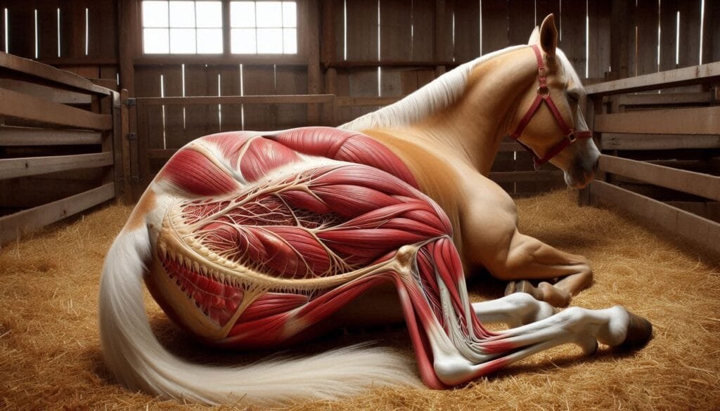
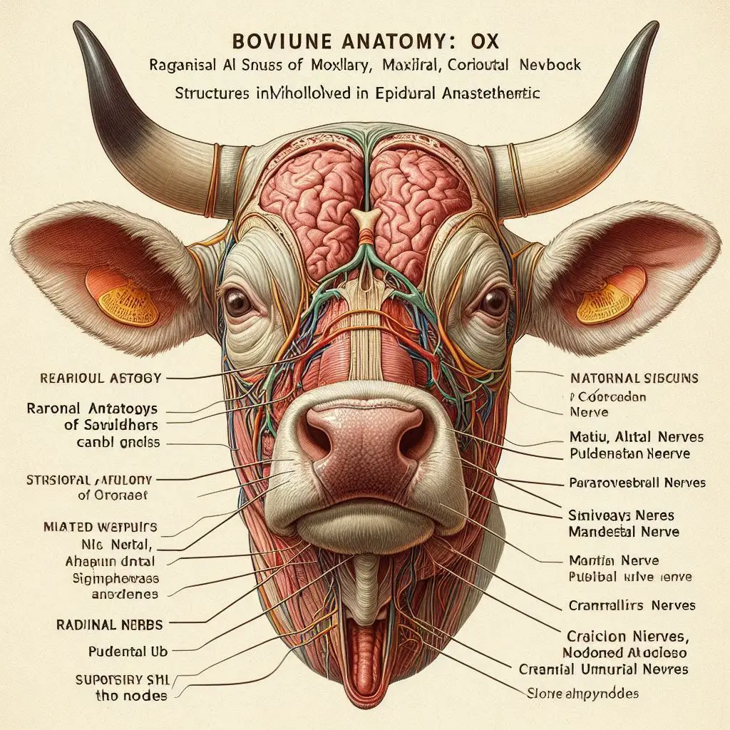
The pudendal nerve is a major nerve in the lumbosacral plexus. It plays a vital role in sensory and motor functions related to the perineum and external genitalia. This nerve is present in various animal species, including dogs, cats, cattle, and sheep. Understanding its anatomy and function helps veterinarians diagnose and treat related conditions.
Importance of the Pudendal Nerve
The pudendal nerve is essential for several reasons:
- It provides sensory input to the external genitalia.
- It controls the muscles involved in urination and defecation.
- It plays a role in reproductive functions.
For more details on the importance of this nerve, you can visit Veterinary Anatomy.
Anatomy of the Pudendal Nerve
Origin and Pathway
The pudendal nerve originates from the sacral plexus. Specifically, it arises from spinal nerves S2 to S4. After its origin, it travels through the greater sciatic foramen. It then re-enters the pelvis through the lesser sciatic foramen.
Branches of the Pudendal Nerve
The pudendal nerve has several important branches:
- Inferior Anal (Rectal) Nerve: This branch innervates the external anal sphincter.
- Perineal Nerve: Supplies sensory innervation to the perineum.
- Dorsal Nerve of the Penis/Clitoris: Provides sensory input to the penis in males and clitoris in females.
For a detailed overview of these branches, check out Pudendal Nerve Anatomy.
Blood Supply
The pudendal nerve receives blood supply from various arteries, including:
- The internal pudendal artery.
- The inferior gluteal artery.
This blood supply is crucial for maintaining nerve function.
Functions of the Pudendal Nerve
Sensory Functions
The pudendal nerve carries sensory information from:
- The skin around the anus.
- The external genitalia.
This sensory input is vital for normal bodily functions like urination and defecation.
Motor Functions
Motor control provided by the pudendal nerve includes:
- Innervation of pelvic floor muscles.
- Control over external anal sphincter and urethral sphincter.
These functions are essential for voluntary control over urination and defecation.
Species Variations
Domestic Animals
In domestic animals such as dogs and cats, studies show that the pudendal nerve’s structure is similar across species. However, there are variations in size and branching patterns based on species differences.
Dogs
In dogs, the pudendal nerve plays a significant role during mating and parturition (giving birth). For more information on canine reproductive anatomy, visit Canine Reproductive Health.
Cats
Cats also exhibit similar anatomical features regarding their pudendal nerve. This nerve contributes significantly to pelvic muscle function during mating.
Farm Animals
Farm animals such as cattle and sheep show adaptations related to their reproductive strategies.
Cattle
In cattle, understanding the pudendal nerve’s role during calving is critical. Proper functioning ensures smooth delivery processes.
Sheep
In sheep, this nerve also plays a role during lambing, affecting both maternal behavior and offspring care.For more insights into reproductive health in farm animals, refer to Farm Animal Reproduction.
Clinical Relevance of the Pudendal Nerve
Understanding the pudendal nerve is crucial for veterinary medicine due to its involvement in various clinical conditions:
Disorders Associated with Pudendal Nerve Dysfunction
Dysfunction of this nerve can lead to several issues:
- Fecal incontinence.
- Urinary retention or incontinence.
These conditions can severely affect an animal’s quality of life.
Surgical Applications
Veterinarians often perform nerve blocks targeting the pudendal nerve during surgical procedures involving:
- The perineum.
- Reproductive organs.
These blocks help manage pain effectively during surgeries.For more information on surgical techniques involving the pudendal nerve, visit Veterinary Surgery Techniques.
Conclusion
The pudendal nerve plays a vital role in both domestic and farm animals by facilitating essential functions related to sensation and motor control in the pelvic region. Understanding its anatomy and function is crucial for improving animal welfare and managing health issues effectively.
More from Veterinary Anatomy:
Pancreas


Responses