
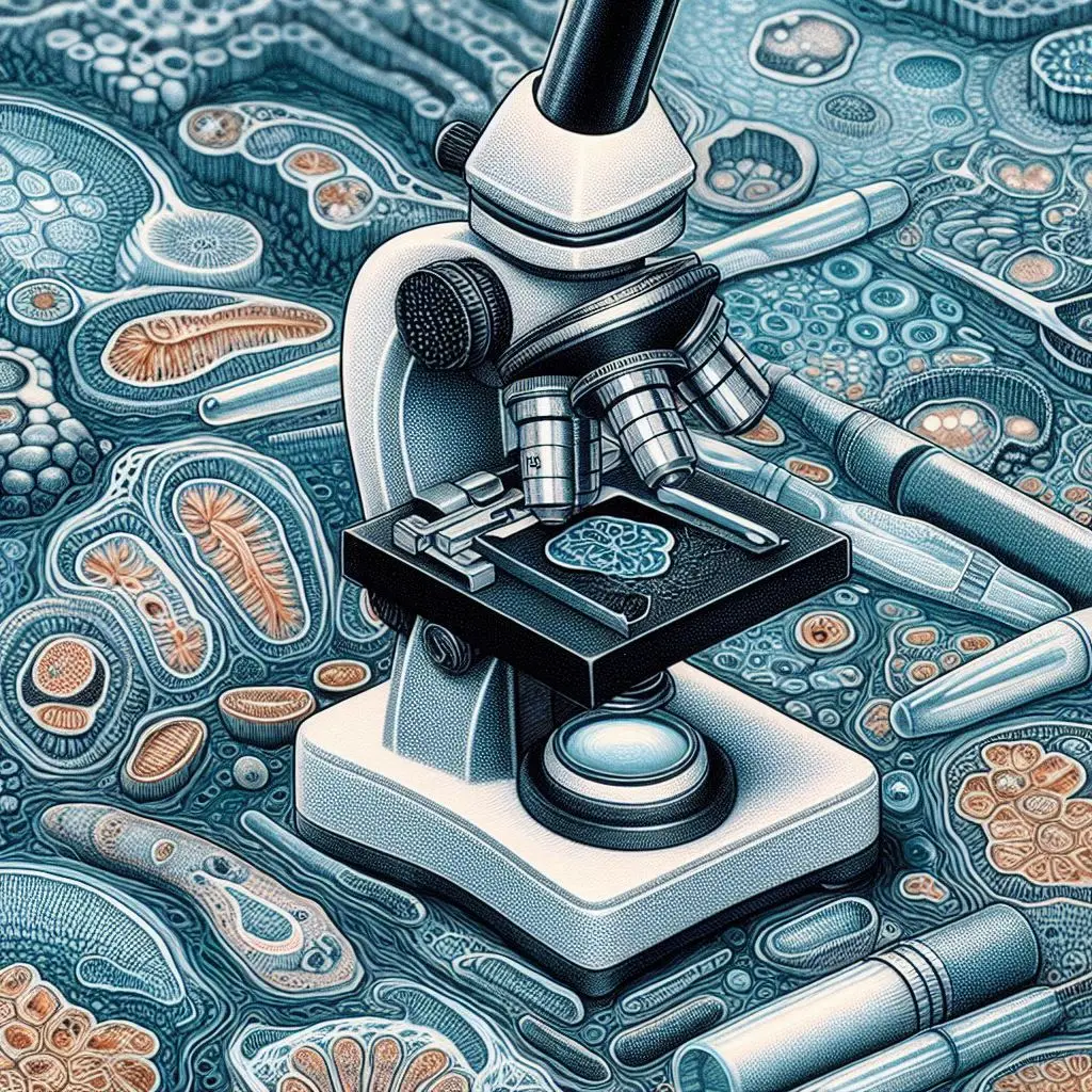
Bright Field Microscopy

Introduction to Bright Field Microscopy
Bright field microscopy is one of the most widely used techniques in optical microscopy. This method allows scientists to observe specimens using transmitted light. Bright field microscopy is particularly effective for examining stained samples, where the specimen appears dark against a bright background. Researchers and educators often rely on this technique for its simplicity and effectiveness, making it a cornerstone in biological and medical research.
What is Bright Field Microscopy?
In this technique, light passes through the specimen and into the objective lens. The contrast arises from the absorption and refraction of light by different parts of the sample. This technique is essential in various fields, including biology, medicine, and materials science.
Historical Context
The roots of bright field microscopy date back to the 17th century when pioneers like Antonie van Leeuwenhoek first observed microorganisms. Over time, advancements in optics and illumination have refined this technique, making it a staple in laboratories worldwide.
How Bright Field Microscopes Work
Basic Principles of Bright Field Microscopy
Bright field microscopes operate on a straightforward principle: illuminating a specimen with light and magnifying the resulting image. Here’s a closer look at how this process works:
- Illumination: A light source, typically a halogen lamp or LED, illuminates the specimen from below.
- Condenser Lens: The condenser focuses light onto the specimen, enhancing illumination.
- Objective Lens: The objective lens collects light from the specimen and magnifies the image.
- Ocular Lens: The eyepiece further magnifies the image for viewing.
Key Components of Bright Field Microscopes
Illuminator
The illuminator is crucial for providing consistent lighting. Modern microscopes often use LED lights for their longevity and brightness.
Condenser
The condenser lens focuses light onto the sample. Adjusting its position can significantly affect image quality.
Objective Lenses
Objective lenses come in various magnifications, typically ranging from 4x to 100x. Each lens has a different numerical aperture (NA), affecting resolution.
Ocular Lens
The ocular lens, or eyepiece, usually provides 10x magnification. Together with the objective lens, it determines total magnification.
Stage
The stage holds the slide securely in place. Some stages are equipped with mechanical controls for precise movement.
Understanding Magnification in Bright Field Microscopy
To calculate total magnification in bright field microscopy, use this formula:
Total Magnification=Magnification of Objective Lens×Magnification of Eyepiece
Total Magnification=Magnification of Objective Lens×Magnification of Eyepiece
For example, if you use a 40x objective lens with a 10x eyepiece, your total magnification will be 400x.
Advantages of Bright Field Microscopy
Simplicity and Ease of Use
One of the main advantages of bright field microscopy is its simplicity. Users can easily set up and operate these microscopes without extensive training. This makes it ideal for educational purposes.
Cost-Effectiveness
Bright field microscopes are generally more affordable than other advanced microscopy techniques like fluorescence or electron microscopy. This makes them accessible to many educational institutions and research labs.
Versatility in Applications
Bright field microscopy can be used across various fields:
- Biology: Observing cellular structures.
- Microbiology: Studying bacteria and other microorganisms.
- Pathology: Analyzing tissue samples for diseases.
- Education: Teaching students about microscopic techniques.
For more information on different models and specifications, visit Olympus’ guide on microscopes.
High Availability of Equipment
Many manufacturers produce bright field microscopes, ensuring that researchers can find suitable equipment easily.
Applications of Bright Field Microscopy
Biological Research Using Bright Field Microscopy
In biological research, bright field microscopy plays a vital role in studying cell morphology and behavior. Researchers can observe living cells or fixed specimens to understand their structure better.
Cell Staining Techniques in Bright Field Microscopy
Staining enhances contrast in bright field microscopy. Common stains include methylene blue and hematoxylin-eosin (H&E). For detailed staining protocols, refer to Nature Protocols.
Medical Diagnostics with Bright Field Microscopy
Pathologists frequently use bright field microscopy to examine tissue samples for abnormalities. This technique helps identify diseases such as cancer by analyzing histological sections.
Histopathology Techniques in Bright Field Microscopy
Histopathology involves preparing thin tissue sections for microscopic examination. Techniques like paraffin embedding and cryosectioning are commonly used in conjunction with bright field microscopy.
Microbial Studies Using Bright Field Microscopy
Microbiologists utilize bright field microscopy to study bacteria and fungi. By applying specific stains, they can differentiate between various types of microorganisms.
Bacterial Staining Methods in Bright Field Microscopy
Gram staining is one common method used to classify bacteria based on their cell wall properties. For more information on Gram staining procedures, check out American Society for Microbiology.
Limitations of Bright Field Microscopy
Low Contrast for Transparent Samples
One significant limitation is that transparent specimens may not provide enough contrast against a bright background. This makes it challenging to visualize certain samples without staining.
Need for Sample Preparation
Many samples require staining or other preparation methods before observation. This process can be time-consuming and may alter the natural state of the specimen.
Limited Depth of Field
Bright field microscopes have a limited depth of field compared to other techniques like confocal microscopy. This can make it challenging to focus on thicker specimens.
Best Practices for Using Bright Field Microscopes
Sample Preparation Techniques
Proper sample preparation is crucial for obtaining high-quality images in bright field microscopy. Here are some tips:
- Use Appropriate Stains: Select stains that enhance contrast without damaging the specimen.
- Thin Sections: Ensure that specimens are thin enough for light to pass through effectively.
- Clean Slides: Always use clean glass slides to avoid artifacts in your images.
Focusing Techniques
Achieving clear images requires careful focusing:
- Start with Low Power: Begin with a low-power objective lens to locate your specimen.
- Adjust Condenser: Fine-tune the condenser height for optimal illumination.
- Use Fine Focus: Once you’ve located your specimen, switch to a higher power and use fine focus for clarity.
Conclusion
Bright field microscopy remains an essential tool in scientific research due to its simplicity and effectiveness in observing biological specimens. While it has limitations regarding contrast and sample preparation, its advantages make it invaluable across various fields such as biology, medicine, and education.
For those interested in exploring further into advanced techniques or alternative methods like fluorescence or electron microscopy, resources such as Microscopy Today provide excellent insights into current trends and technologies in microscopy.
For more pearls of Vets Wisdom:
Current Status Of Egg Production in India


Your point of view caught my eye and was very interesting. Thanks. I have a question for you.
I don’t think the title of your article matches the content lol. Just kidding, mainly because I had some doubts after reading the article.