Comparative Histology of the Brain
Introduction to Brain Histology
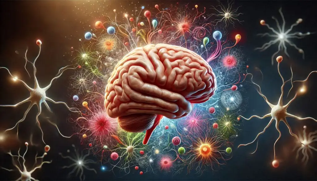
Histology is the study of tissues at a microscopic level. The brain’s histological organization reveals much about its function. It consists of two main types of tissue: gray matter and white matter. Each type plays a unique role in brain activity.
What is Gray Matter?
Gray matter contains neuronal cell bodies, dendrites, and glial cells. It is primarily involved in processing information. The cerebral cortex, which is the outer layer of the brain, is rich in gray matter. This area is crucial for sensory perception, cognition, and motor control.
Understanding White Matter
White matter consists mainly of myelinated axons. These axons connect different regions of the brain and spinal cord. The myelin sheath gives white matter its lighter appearance. This tissue facilitates rapid signal transmission between neurons.
Major Cell Types in the Brain
The brain contains several cell types that contribute to its function:
Neurons
Neurons are the primary functional units of the brain. They transmit signals throughout the nervous system. There are various types of neurons, each with specific roles:
- Pyramidal Neurons: These are found in the cerebral cortex and are characterized by their pyramid-shaped cell bodies.
- Interneurons: These neurons connect other neurons within specific regions of the brain.
For more on neuron types and their functions, visit Neuroscience Online.
Glial Cells
Glial cells support and protect neurons. They play several critical roles:
- Astrocytes: These star-shaped cells help maintain the blood-brain barrier (BBB) and supply nutrients to neurons.
- Oligodendrocytes: Responsible for producing myelin in the central nervous system (CNS).
- Microglia: Act as immune cells in the CNS, responding to injury or disease.
To learn more about glial cells, refer to this article on The Role of Glia in Brain Function.
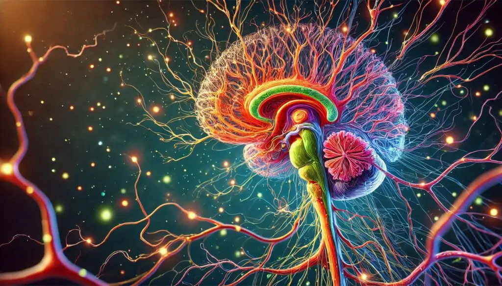
Histological Features of Different Brain Regions
The brain has several distinct regions, each with unique histological features.
Cerebral Cortex
The cerebral cortex is divided into six layers (neocortex), each with specific functions:
Layer 1: Molecular Layer
This layer contains few neurons but many dendrites from deeper layers.
Layer 2: External Granular Layer
This layer has small pyramidal neurons and many granule cells.
Layer 3: External Pyramidal Layer
It consists mainly of medium-sized pyramidal neurons that project to other cortical areas.
Layer 4: Internal Granular Layer
This layer receives sensory input from the thalamus and has numerous granule cells.
Layer 5: Internal Pyramidal Layer
It contains large pyramidal neurons that send outputs to subcortical structures.
Layer 6: Multiform Layer
This layer contains various neuron types that project to different areas.
For detailed insights into cortical layers, check out The Human Connectome Project.
Choroid Plexus
The choroid plexus is a specialized tissue found within the brain’s ventricles. It produces cerebrospinal fluid (CSF), which cushions and protects the brain. The choroid plexus consists of a highly vascularized stroma lined by ependymal cells.
For more information on CSF production and function, visit Cerebrospinal Fluid Dynamics.
Blood-Brain Barrier (BBB)
The blood-brain barrier is crucial for protecting the brain from harmful substances while allowing essential nutrients to pass through. It consists of endothelial cells connected by tight junctions, astrocytic processes, and pericytes.
To understand how the BBB functions, read more at Blood-Brain Barrier Overview.
Functional Implications of Brain Histology
The histological organization directly impacts how the brain functions:
Efficient Signal Transmission
The arrangement of gray and white matter allows for efficient communication between different parts of the brain. Myelinated axons in white matter enable faster signal transmission compared to unmyelinated fibers.
Neuronal Health and Support
Glial cells play a vital role in maintaining neuronal health. They provide support, nutrients, and protection against injury or disease.
Protection from Toxins
The blood-brain barrier protects neural tissue from potentially harmful substances while allowing necessary exchanges with blood. This selective permeability is crucial for maintaining a stable environment for neuronal function.
Comparative Histology Across Species
Understanding how different species’ brains are organized can provide insights into evolution and function.
Human vs. Animal Brains
Humans have a highly developed cerebral cortex compared to many animals. This development supports advanced cognitive functions such as reasoning and problem-solving.
For a deeper understanding of comparative neuroanatomy, check out Comparative Neuroanatomy.
Primates vs. Rodents
Primates generally have larger brains relative to body size than rodents. The increased complexity in primate brains supports more sophisticated social behaviors and learning capabilities.
To explore this topic further, visit Primate Neuroanatomy.
Conclusion
In summary, understanding the comparative histology of the brain offers valuable insights into its complex structure and function. The interplay between different cell types and tissue structures underlies both normal operations and pathological conditions within the central nervous system.
More from Veterinary Anatomy:
Microscopy Techniques


Responses