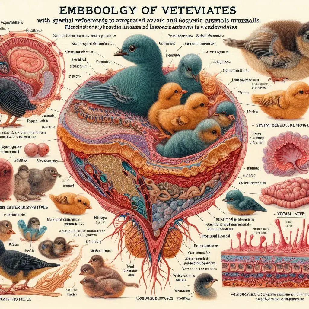
Endotheliochorial Placenta

Introduction to the Endotheliochorial Placenta
The endotheliochorial placenta is a fascinating topic in the study of mammalian reproduction. This type of placenta is particularly notable in carnivores, such as dogs and cats. Understanding its structure and function can provide insights into how these animals nourish their young during gestation.
What is a Placenta?
Before diving into the specifics of the endotheliochorial placenta, it’s essential to understand what a placenta is. The placenta is an organ that develops during pregnancy. It connects the developing fetus to the uterine wall, facilitating nutrient uptake, waste elimination, and gas exchange via the mother’s blood supply.
For more information on placental functions, you can visit Healthline.
Structure of the Endotheliochorial Placenta
Key Components
The endotheliochorial placenta consists of several layers that separate maternal and fetal blood:
- Chorionic Epithelium: This layer comes from the fetal tissue.
- Endothelial Cells: These cells line the fetal blood vessels.
- Maternal Endometrial Epithelium: This layer is part of the mother’s uterine lining.
In this type of placenta, there are fewer layers compared to other types, such as epitheliochorial or hemochorial placentas.
Comparison with Other Placental Types
- Epitheliochorial Placenta: Found in pigs and cows, this type retains all six tissue layers between maternal and fetal blood.
- Hemochorial Placenta: Seen in humans and rodents, this type allows direct contact between chorionic tissue and maternal blood.
For a detailed comparison of these placental types, check out Wikipedia.
Layers in Detail
The endotheliochorial placenta has four main layers:
- Chorionic Epithelium
- Chorionic Connective Tissue
- Maternal Endothelium: This layer consists of maternal blood vessels.
- Maternal Blood: In endotheliochorial placentas, maternal blood is close to fetal tissues.
This structure allows for efficient nutrient transfer while maintaining some separation between maternal and fetal circulations.
Function of the Endotheliochorial Placenta
Nutrient Transfer
One of the primary functions of any placenta is to facilitate nutrient transfer from mother to fetus. In carnivores with an endotheliochorial placenta, this process is efficient due to the reduced number of tissue layers.
Mechanism of Nutrient Transfer
Nutrients from maternal blood pass through the chorionic epithelium into fetal circulation. This process involves simple diffusion and active transport mechanisms.
Gas Exchange
Gas exchange is another critical function. Oxygen passes from maternal blood to fetal blood while carbon dioxide moves in the opposite direction. The close proximity of maternal blood vessels enhances this exchange.
For more insights into how gas exchange occurs in placentas, refer to ScienceDirect.
Hormonal Functions
The placenta also plays a role in hormone production during pregnancy. Hormones like progesterone and estrogen are crucial for maintaining pregnancy and preparing for childbirth.
Evolutionary Significance
Adaptations in Carnivores
The endotheliochorial placenta represents an evolutionary adaptation that allows carnivores to efficiently nourish their young. This efficiency is vital for species survival, especially in environments where resources may be scarce.
Evolutionary Trends
The trend towards more invasive placentation types reflects an evolutionary strategy to enhance fetal development. As species evolved, different placental structures emerged to meet specific reproductive needs.
For further reading on evolutionary biology related to placentation, visit National Geographic.
Advantages of Endotheliochorial Placenta
Efficient Nutrient Transfer
The reduced number of layers allows for faster nutrient transfer compared to other types of placentas. This efficiency can lead to healthier offspring with better chances of survival.
Enhanced Gas Exchange
With fewer barriers between maternal and fetal blood, gas exchange becomes more effective. This feature is especially important for carnivores that may have high metabolic demands.
Flexibility in Gestation Periods
Carnivores often have varied gestation periods based on environmental conditions. The endotheliochorial structure provides flexibility that can adapt to these changing conditions.
Disadvantages of Endotheliochorial Placenta
Increased Risk of Maternal-Fetal Blood Mixing
One potential downside is the increased risk of maternal-fetal blood mixing. This situation can lead to complications if there are incompatible blood types between mother and fetus.
Limited Maternal Tissue Protection
While the close contact enhances nutrient transfer, it also means less maternal tissue protection for the fetus compared to more layered placentas like epitheliochorial ones.
Examples of Animals with Endotheliochorial Placentas
Dogs (Canis lupus familiaris)
Dogs are one of the most studied species with an endotheliochorial placenta. Their gestation period lasts about 63 days, during which efficient nutrient transfer is crucial for puppy development.
For more information about dog reproduction, check out American Kennel Club.
Cats (Felis catus)
Cats also possess an endotheliochorial placenta. Their gestation period ranges from 58 to 67 days. Like dogs, cats benefit from this efficient placental structure during pregnancy.
To learn more about cat reproduction, visit PetMD.
Conclusion
The endotheliochorial placenta plays a vital role in the reproductive success of carnivorous mammals like dogs and cats. Its unique structure allows for efficient nutrient transfer and gas exchange while presenting some risks associated with maternal-fetal interactions.
More from Veterinary Anatomy:
Epithelial Tissue


Responses