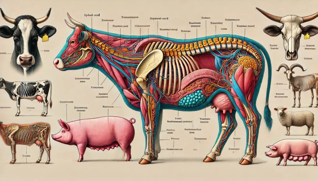
Epidural Anesthesia in Cattle: Techniques and Anatomy

Introduction
Epidural anesthesia is a valuable technique in veterinary medicine, particularly for cattle. This method provides effective pain relief during surgical procedures and assists in managing various medical conditions. Understanding the anatomy involved and the techniques used for epidural anesthesia in cattle is crucial for veterinarians and animal handlers alike. In this article, we will explore the relevant structures, techniques, and best practices for performing epidural anesthesia in bovines.
Understanding the Anatomy
The Spinal Column
The spinal column of cattle consists of several vertebrae. Cattle have:
- 7 cervical vertebrae
- 13 thoracic vertebrae
- 6 lumbar vertebrae
- 5 sacral vertebrae (fused into the sacrum)
- 18-20 coccygeal vertebrae
This structure is vital for understanding where to administer the epidural injection.
The Epidural Space
The epidural space is the area surrounding the dura mater of the spinal cord. It extends from the foramen magnum at the base of the skull to the sacrococcygeal ligament. This space is crucial for the administration of anesthetic agents.
- Boundaries of the Epidural Space:
- Posteriorly: Ligamentum flavum
- Laterally: Pedicles and intervertebral foramina
- Anteriorly: Posterior longitudinal ligament
Nerve Supply
The nerve supply to the pelvic and abdominal regions is essential for understanding the effects of epidural anesthesia.
- Preganglionic sympathetic fibers originate from the spinal cord levels T1 to L2.
- Parasympathetic fibers come from S2 to S4. These fibers control functions in the bladder, large intestine, and rectum.
Key Landmarks for Injection
Identifying the correct landmarks is vital for successful epidural anesthesia. The most common sites for injection in cattle are:
- Sacrococcygeal intervertebral space (S5-Co1)
- First intercoccygeal intervertebral space (Co1-Co2)
The transverse processes of lumbar vertebrae L2 to L5 serve as additional landmarks for paravertebral nerve blocks.
Techniques for Epidural Anesthesia
Performing epidural anesthesia requires a clear understanding of the technique. Here’s a step-by-step guide:
1. Preparation
Before starting, gather all necessary equipment:
- A 1.5-inch, 18-gauge needle
- Sterile anesthetic solution (e.g., lidocaine or bupivacaine)
- Antiseptic solution for skin preparation
- Gloves and other personal protective equipment
2. Positioning the Animal
Position the cattle comfortably. It is often best to have the animal standing or in lateral recumbency. Ensure that the tail is accessible for the injection.
3. Identifying the Injection Site
Locate the sacrococcygeal space or the first intercoccygeal space. Palpate the area to ensure proper positioning.
4. Administering the Injection
- Insert the needle at a 45-degree angle toward the tail.
- Keep the bevel of the needle facing upward. This positioning helps the anesthetic diffuse more effectively.
- Use the hanging drop technique. Place a drop of lidocaine in the hub of the needle. When you reach the epidural space, the drop will be sucked into the needle.
5. Injecting the Anesthetic
Once you confirm you are in the epidural space, slowly inject the anesthetic solution. Monitor the animal closely for any signs of adverse reactions.
6. Post-Procedure Care
After administering the epidural, keep the animal calm and monitor its recovery. Ensure that the anesthesia is effective and that the animal is comfortable.
Considerations and Best Practices
Dosage and Drug Selection
Choosing the right drug and dosage is crucial for effective anesthesia. Common agents include lidocaine and bupivacaine. The dosage will depend on the size of the animal and the specific procedure. Always refer to veterinary guidelines for appropriate dosages.
Monitoring the Animal
After the procedure, closely monitor the animal for any signs of complications. Look for:
- Changes in behavior
- Signs of pain or discomfort
- Difficulty in movement
Complications
While epidural anesthesia is generally safe, complications can occur. Possible complications include:
- Inadvertent injection into the subarachnoid space
- Hematoma formation
- Infection at the injection site
Be prepared to manage these complications promptly.
Conclusion
Epidural anesthesia is a vital technique in veterinary medicine for cattle. Understanding the anatomy involved and mastering the injection technique can lead to successful outcomes. With proper preparation, monitoring, and care, veterinarians can provide effective pain relief for bovines undergoing surgical procedures.
For more pearls of Vets Wisdom:
https://wiseias.com/partitioning-of-food-energy-within-animals/


Responses