
Freezing Microtomes

Introduction to Freezing Microtomes
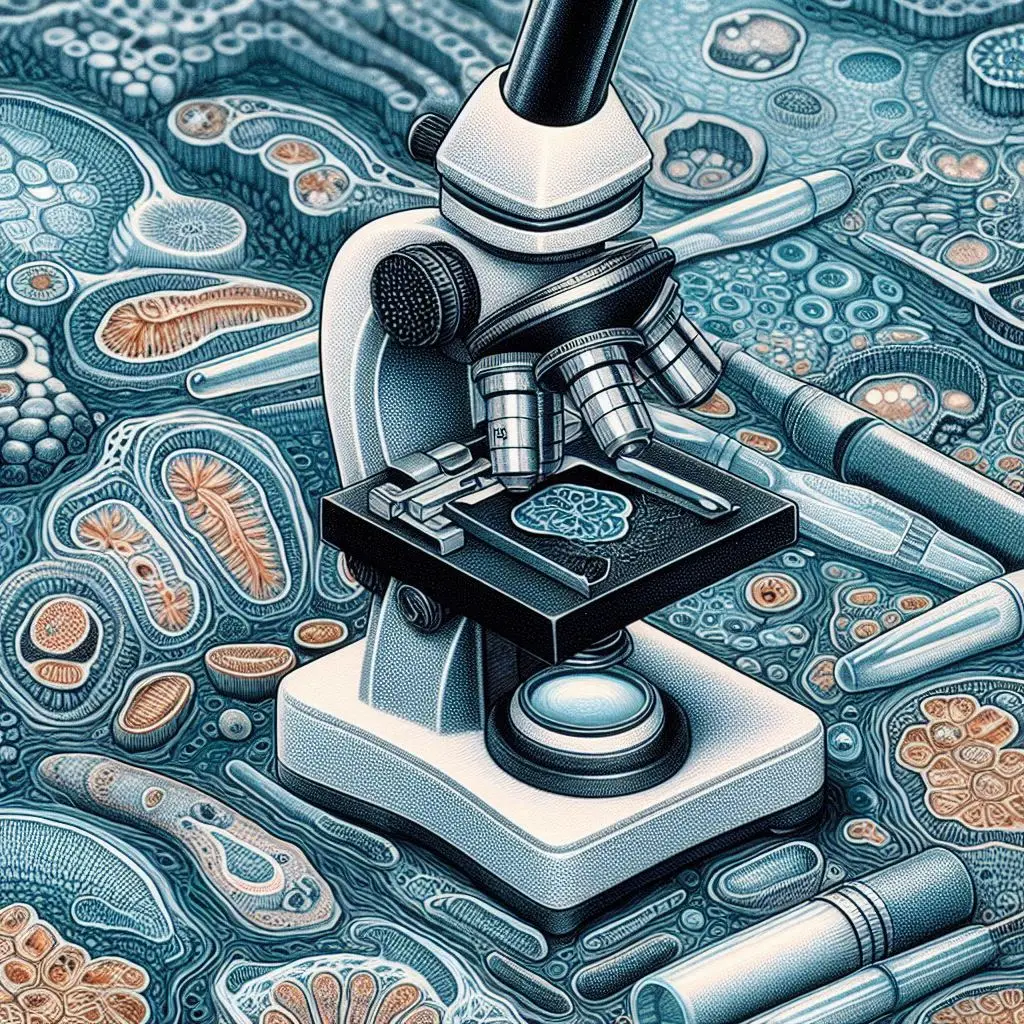
Freezing microtomes, also known as cryostats, are essential tools in histology and pathology. These instruments allow researchers and medical professionals to cut thin sections of frozen tissue samples. By using a freezing microtome, the structural integrity of the tissues is preserved, making it easier to analyze them under a microscope. In this article, we will explore how freezing microtomes work, their main components, and their applications in various fields.
What is a Freezing Microtome?
A freezing microtome is a specialized instrument designed to slice frozen biological specimens into thin sections. It operates at low temperatures to maintain the integrity of the tissue. By using a freezing microtome, pathologists can prepare samples for immediate examination. This is crucial for accurate diagnoses and research. For more information on the importance of freezing microtomes in histology, check out Nature Reviews.
Importance of Freezing Microtomes in Histology
Histology is the study of tissues at a microscopic level. Freezing microtomes play a critical role in this field by allowing for rapid processing of samples. They help preserve cellular structures that might otherwise degrade during traditional fixation methods. For detailed insights into histology techniques, you can visit ScienceDirect.
How Freezing Microtomes Work
The Cutting Process
The cutting process involves several steps:
- Freezing the Sample: The tissue sample is first frozen using liquid nitrogen or a refrigeration unit.
- Sectioning: The frozen specimen is then placed in the microtome for slicing.
- Collection: The thin sections are collected for further analysis.
This method ensures that the cellular details remain intact, which is vital for accurate diagnosis.
Temperature Control Mechanism
Temperature control is crucial in freezing microtomes. The freezing chamber maintains low temperatures throughout the cutting process. This prevents the tissue from thawing and preserves its structure. For more on temperature regulation in laboratory settings, check out Lab Manager.
Main Components of Freezing Microtomes
Understanding the components of freezing microtomes helps clarify their operation.
1. Freezing Chamber
The freezing chamber houses the specimen during sectioning. It includes advanced refrigeration systems that maintain optimal low temperatures.
2. Sectioning Mechanism
The sectioning mechanism consists of:
- Blade: A sharp blade slices through frozen tissues.
- Advance/Retract Mechanism: This allows precise control over section thickness.
3. Specimen Holder
The specimen holder secures the frozen sample during cutting. It ensures stability and accuracy while slicing.
4. Temperature Control System
This system regulates the temperature within the freezing chamber. It uses sensors to maintain consistent conditions throughout the process.
5. Knife Holder and Blade
The knife holder supports the cutting blade, which must be sharp enough to slice through frozen tissue effectively.
6. Defrosting Mechanism
Some models include a defrosting mechanism that manages ice buildup on components, ensuring smooth operation.
7. Display and Controls
User interfaces allow monitoring and adjusting parameters like temperature and section thickness.
8. Illumination System
An illumination system provides light to help visualize the specimen during sectioning.
9. Waste Collection System
This system collects debris generated during cutting for easy disposal.
Applications of Freezing Microtomes
Freezing microtomes are invaluable in various fields:
1. Histology and Pathology
In histology and pathology, freezing microtomes prepare specimens for immunohistochemistry and in situ hybridization studies. These techniques rely on high-quality tissue sections for accurate results.
2. Research Laboratories
Research laboratories use freezing microtomes for rapid processing of samples critical in time-sensitive studies. They enable scientists to analyze tissues quickly without compromising quality.
3. Biobanks
Biobanks store biological samples for future analysis. Freezing microtomes help preserve these samples by allowing precise sectioning without damaging cellular structures. For more information on biobanking practices, visit Biobanking.org.
Advantages of Using Freezing Microtomes
Using freezing microtomes offers several advantages:
- Rapid Processing: Samples can be prepared quickly, which is crucial in clinical settings.
- Preservation of Cellular Integrity: The freezing process maintains cellular structures that might degrade with traditional methods.
- Versatility: They can be used for various types of tissues, including those that are delicate or difficult to handle.
- Improved Diagnostic Accuracy: High-quality sections lead to better diagnostic outcomes in pathology.
Challenges and Considerations
While freezing microtomes have many benefits, there are also challenges:
1. Skill Requirement
Operating a freezing microtome requires skill and training. Proper technique is essential for producing high-quality sections.
2. Equipment Maintenance
Regular maintenance is necessary to ensure optimal performance of the equipment.
3. Cost Factors
Freezing microtomes can be expensive investments for laboratories, which may limit accessibility for some institutions.
Conclusion
Freezing microtomes are vital tools in histology and pathology, enabling precise sectioning of frozen tissues while preserving their integrity. Understanding how they work and their components can enhance their effective use in research and clinical settings.
For more pearls of Vets Wisdom:


Thanks for sharing. I read many of your blog posts, cool, your blog is very good.
I don’t think the title of your article matches the content lol. Just kidding, mainly because I had some doubts after reading the article. https://www.binance.info/en-IN/register?ref=UM6SMJM3
Your point of view caught my eye and was very interesting. Thanks. I have a question for you.
Your article helped me a lot, is there any more related content? Thanks! https://accounts.binance.info/en/register?ref=JHQQKNKN