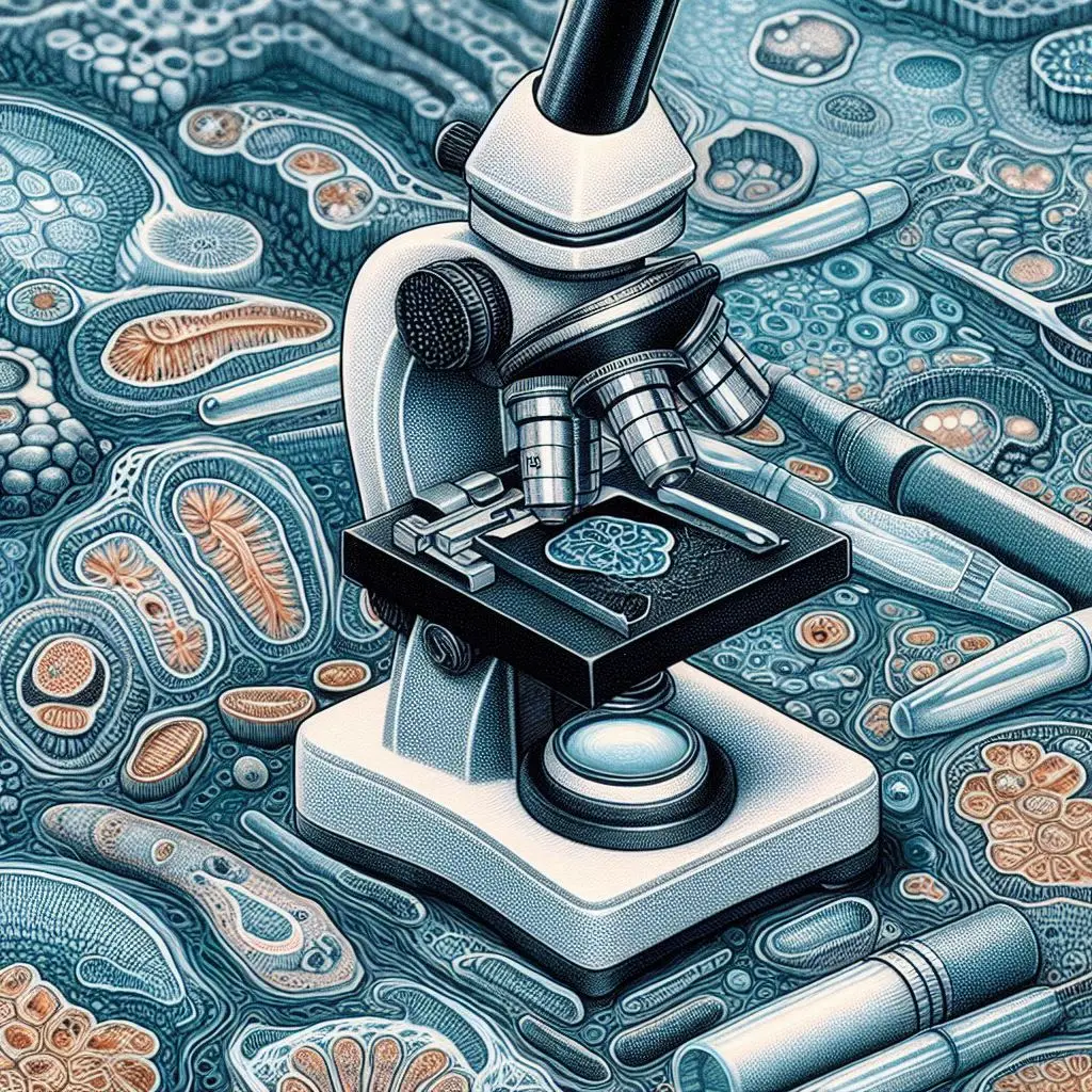
Freezing Microtomes: The Essential Tool for Cutting Thin Tissue Sections

Freezing microtomes are specialized instruments crucial for preparing thin tissue sections. They allow precise sectioning of biological samples without damaging tissue structure. This article explores the functionality, key features, and applications of freezing microtomes. We will also discuss the different types available.
Functionality
Freezing microtomes operate at temperatures between -60°C and 0°C. They use liquid nitrogen or cryogenic coolants to maintain these low temperatures. Rapid freezing minimizes ice crystal formation. This process protects cellular integrity and prevents artifacts that can compromise sample quality.The freezing microtome has a deep-cooled platform. Technicians place the sample on this platform to freeze it. After cutting each section, the platform raises to the desired thickness. This allows for sections ranging from 5 μm to 40 μm.
Key Features
Temperature Control
Freezing microtomes maintain a controlled environment for quick freezing. This preservation of molecular structures is essential for preventing ice crystal formation.
Cutting Mechanism
These devices include adjustable cutting blades. Technicians can slice sections of varying thicknesses, typically between 5 μm and 40 μm. They collect these sections for further analysis, such as histological staining or electron microscopy.
Applications
Freezing microtomes are widely used in histopathology. They prepare tissue samples from biopsies. Additionally, they find applications in various industries, slicing materials like textiles and food products.
Types of Freezing Microtomes
Standard Freezing Microtomes
Standard freezing microtomes feature a platform that freezes the sample directly. They allow for cutting thicker sections.
Cryostats
Cryostats are advanced systems that maintain a consistent low temperature in a chamber. They provide a controlled environment for sectioning. These devices can produce sections ranging from 10 μm to 30 μm.
Ultracryotomes
Ultracryotomes create ultrathin sections for transmission electron microscopy. They can achieve thicknesses on the nanometer scale. This capability enables detailed ultrastructural studies.
Advantages of Freezing Microtomes
- Better Molecular Preservation: Rapid freezing helps maintain molecular integrity. This preservation is crucial for techniques requiring molecular detection.
- Versatility in Section Thickness: Frozen samples can be sectioned into thin (5 μm to 15 μm), thick (dozens of μm), and ultrathin (nanometers) sections for specific applications.
- Suitability for Various Tissues: Freezing microtomes produce good-quality sections from many tissues. However, they may struggle with mineralized tissues like bone or highly keratinized tissues like horns.
- Faster Sample Preparation: Technicians can quickly freeze and section unfixed material, such as biopsies. This process eliminates the need for fixation and embedding.
Sample Preparation
To preserve tissue integrity during freezing, it is crucial to prevent large ice crystals from forming. There are two main methods for achieving this:
- Fast and Deep Freezing: Rapidly freezing the sample with liquid nitrogen minimizes ice crystal formation.
- Antifreeze Immersion: If the sample does not need immediate cutting, technicians can fix it and immerse it in an antifreeze solution. This solution contains sucrose and glycerol, preventing large ice crystals while allowing for cellular ultrastructure studies.
Conclusion
Freezing microtomes are essential tools in medical and industrial settings. They enable high-quality sample preparation for microscopic examination while maintaining biological material integrity. By understanding the functionality, features, and applications of these devices, researchers and technicians can optimize their tissue sectioning processes. This knowledge leads to valuable insights from their samples.
For more pearls of Vets Wisdom:
https://wiseias.com/partitioning-of-food-energy-within-animals/


Responses