Hip Dislocation Surgery in Large Animals
Surgical Management of Hip Dislocation in Large Animals
Hip dislocation, also called hip luxation, occurs when the femoral head moves out of the acetabulum. It is a severe orthopedic condition in large animals like cattle and horses. Immediate intervention is necessary to prevent complications such as permanent joint damage, chronic pain, or lameness. In many cases, hip dislocation results from high-impact trauma, excessive force during handling, or underlying musculoskeletal weakness.
Causes and Clinical Signs
Common Causes of Hip Dislocation
Hip dislocation in large animals is often caused by external trauma or an internal predisposition. The following are some of the primary causes:
- Trauma: Sudden falls, slipping on wet or uneven surfaces, collisions, or excessive force during assisted calving can result in hip luxation.
- Congenital hip dysplasia: Some large animals, particularly cattle breeds, may have a genetic predisposition to weak hip joints, increasing the risk of dislocation.
- Weak ligaments and muscle injuries: Over time, ligaments and muscles supporting the hip joint may weaken due to poor nutrition, lack of exercise, or excessive weight-bearing, making dislocation more likely.
- Excessive force during handling: Improper restraint during medical procedures, rough transportation, or sudden jerks in confined spaces may lead to joint displacement.
- Joint infections or inflammation: Chronic infections like septic arthritis weaken the hip joint, making it susceptible to dislocation.
Recognizing the Signs of Hip Dislocation
Detecting hip luxation early can significantly improve recovery chances. Some common clinical signs include:
- Severe lameness and visible pain: The affected animal may refuse to bear weight on the injured limb.
- Abnormal limb positioning: The leg may be rotated outward or inward, depending on the type of dislocation.
- Muscle atrophy: If left untreated, muscles in the affected limb may shrink due to disuse.
- Swelling and joint instability: Palpation of the hip region may reveal abnormal movement or a misaligned femoral head.
- Difficulty standing or walking: Severe cases may result in an inability to rise or maintain balance.
Diagnosis and Initial Assessment
Clinical Examination
Veterinarians begin with a thorough physical examination to check for hip dislocation. This includes:
- Observing the animal’s stance and gait
- Palpating the hip region to assess joint stability
- Performing limb manipulation tests
- Evaluating pain response and potential nerve damage
Radiographic Confirmation
X-rays are the gold standard for confirming hip luxation and ruling out fractures. They provide a clear image of the femoral head’s displacement. Sedation is often necessary for a proper radiographic examination, as large animals may struggle during positioning (American Veterinary Medical Association).
Surgical and Non-Surgical Treatment Options
Closed Reduction: Non-Surgical Approach
If the dislocation is recent and there are no fractures, a closed reduction can be attempted. The steps include:
- Administering sedation or general anesthesia to relax the muscles and prevent pain.
- Manual manipulation of the femoral head back into the hip socket using traction and rotation techniques.
- Stabilizing the hip joint with a bandage, sling, or hobbles to prevent re-dislocation.
- Monitoring joint stability through post-reduction X-rays.
Closed reduction works best in acute cases but may not be suitable if ligaments or joint capsules are severely damaged (Merck Veterinary Manual).
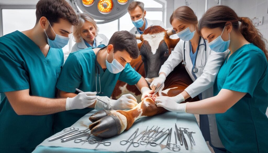
Open Reduction: Surgical Treatment
If closed reduction fails or if the dislocation is chronic, surgery is required. The primary surgical techniques include:
Capsulorrhaphy
- Involves suturing the joint capsule to restore stability.
- Works well for cases with minimal soft tissue damage.
Toggle Pin Fixation
- A surgical implant secures the femoral head within the acetabulum.
- Suitable for severe ligament tears or chronic dislocations.
Femoral Head Ostectomy (FHO)
- The femoral head is surgically removed to eliminate pain.
- Recommended for non-weight-bearing animals or cases where joint repair is impossible.
Postoperative Care and Recovery
Pain Management and Support
Pain control is crucial for recovery. Veterinarians use NSAIDs, opioids, and local anesthetics to manage discomfort. Proper stall rest and restricted movement also aid healing.
Rehabilitation and Physiotherapy
- Controlled weight-bearing exercises prevent muscle atrophy.
- Hydrotherapy is excellent for muscle strengthening.
- Cold therapy reduces swelling in the early postoperative period.
- Massage therapy enhances circulation and prevents stiffness.
A structured physiotherapy plan significantly improves joint mobility (University of Minnesota Veterinary Medicine).
Monitoring for Complications
Key risks include:
- Reluxation if the joint remains unstable.
- Joint infections post-surgery.
- Chronic muscle atrophy if rehabilitation is delayed.
Regular follow-up radiographs and veterinary check-ups are essential to track progress.
Preventing Hip Dislocation in Large Animals
Proper Handling and Housing
Preventative strategies include:
- Installing non-slip flooring in barns and transport vehicles.
- Avoiding overcrowding in stalls and pens.
- Training handlers in low-stress animal management techniques.
Nutritional Support
- Maintaining optimal calcium and phosphorus levels to support bone health.
- Providing joint supplements such as glucosamine and chondroitin.
Balanced nutrition reduces the risk of joint issues (National Animal Health Monitoring System).
Conclusion
Early detection and prompt surgical management of hip dislocation in large animals lead to better outcomes. Proper treatment, rehabilitation, and preventive care can significantly reduce recurrence and improve mobility. If you suspect hip dislocation in your animal, consult a veterinary specialist immediately to determine the best course of action.
For more pearls of Vets Wisdom:
Comminuted Fractures in Animals

Responses