Surface Anatomy of Visceral Organs in the Thoracic, Abdominal and Pelvic Cavities
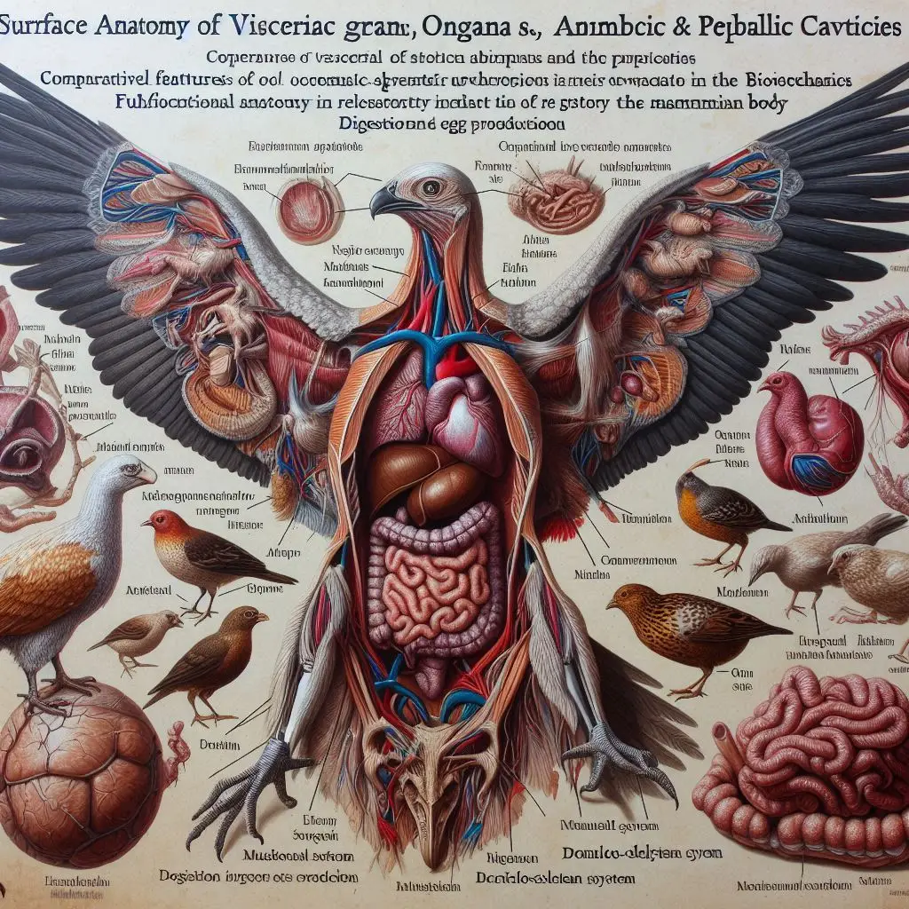
This comprehensive guide will explore the surface anatomy of the visceral organs located within the thoracic, abdominal and pelvic cavities. We’ll dive deep into the key structures and relationships of these vital organs to provide a thorough understanding of their surface anatomy.
Thoracic Cavity
The thoracic cavity sits superior to the diaphragm and is divided into several important compartments:
Pleural Cavities
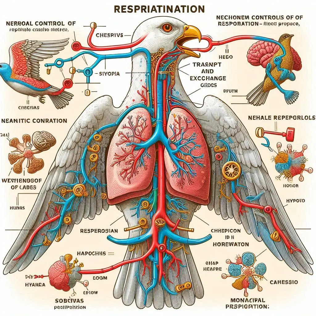
The pleural cavities contain the lungs, which are essential for respiration. Each lung is surrounded by a pleural membrane, allowing for expansion and contraction during breathing.
Mediastinum
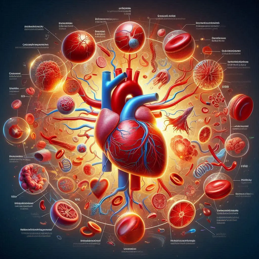
The mediastinum is the central compartment that houses critical structures like the heart, trachea, esophagus, and major blood vessels. The heart resides within the pericardial cavity, positioned between the lungs and protected by the sternum anteriorly and vertebral column posteriorly.
Abdominal Cavity
The abdominal cavity lies inferior to the thoracic cavity and superior to the pelvic cavity. It contains a diverse array of organs primarily associated with digestion and metabolism:
Digestive Organs
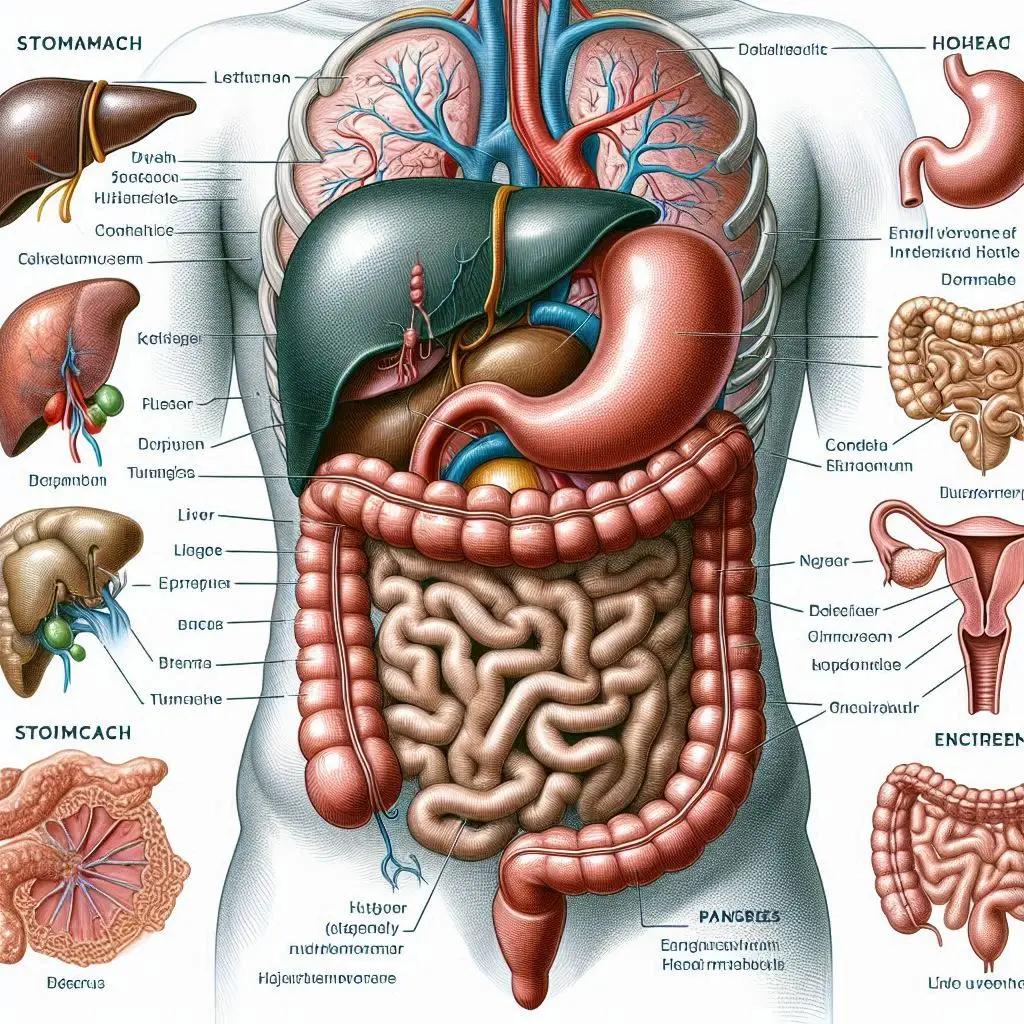
The stomach, small intestine (duodenum, jejunum, ileum), and large intestine (cecum, colon, rectum) are crucial for food processing and nutrient absorption.
Accessory Organs
The liver, gallbladder, and pancreas play vital roles in digestion and metabolism. The liver produces bile, the gallbladder stores it, and the pancreas produces digestive enzymes and hormones.
Other Organs
The spleen (part of the immune system) and kidneys (part of the urinary system) are also located in this cavity. The kidneys are retroperitoneal, situated behind the peritoneum, while the spleen is located in the left upper quadrant. The abdominal cavity is lined by the peritoneum, which consists of two layers: the parietal peritoneum lining the cavity wall and the visceral peritoneum covering the organs. This membrane creates a potential space, the peritoneal cavity, which contains serous fluid to reduce friction between organs during movement.
Pelvic Cavity
The pelvic cavity is situated inferior to the abdominal cavity and is bounded by the pelvic girdle. It contains:
Reproductive Organs
In females, this includes the uterus, ovaries, and fallopian tubes. In males, it includes the prostate and seminal vesicles.
Urinary System
The urinary bladder and urethra are located here, responsible for urine storage and excretion.
Rectum
The final section of the large intestine, leading to the anus, is also situated in the pelvic cavity.
Relationships Between Organs
Understanding the surface anatomy of the visceral organs in the thoracic, abdominal, and pelvic cavities is crucial for medical professionals. This knowledge aids in diagnosing and treating various medical conditions, as well as performing surgical procedures effectively. Each cavity serves distinct functions, housing organs that are intricately connected through systems of blood supply, innervation, and lymphatic drainage, contributing to the overall homeostasis of the body.
Thoracic Cavity Relationships
The pleural cavities contain the lungs, which are essential for respiration. Each lung is surrounded by a pleural membrane, allowing for expansion and contraction during breathing. The mediastinum houses critical structures like the heart, trachea, esophagus, and major blood vessels. The heart resides within the pericardial cavity, positioned between the lungs and protected by the sternum anteriorly and vertebral column posteriorly.
Abdominal Cavity Relationships
The abdominal cavity contains a diverse array of organs primarily associated with digestion and metabolism. The stomach, small intestine (duodenum, jejunum, ileum), and large intestine (cecum, colon, rectum) are crucial for food processing and nutrient absorption. The liver, gallbladder, and pancreas play vital roles in digestion and metabolism. The spleen (part of the immune system) and kidneys (part of the urinary system) are also located in this cavity. The kidneys are retroperitoneal, situated behind the peritoneum, while the spleen is located in the left upper quadrant. The abdominal cavity is lined by the peritoneum, which consists of two layers: the parietal peritoneum lining the cavity wall and the visceral peritoneum covering the organs. This membrane creates a potential space, the peritoneal cavity, which contains serous fluid to reduce friction between organs during movement.
Pelvic Cavity Relationships
The pelvic cavity contains reproductive organs, the urinary system, and the rectum. In females, this includes the uterus, ovaries, and fallopian tubes. In males, it includes the prostate and seminal vesicles. The urinary bladder and urethra are located here, responsible for urine storage and excretion. The final section of the large intestine, leading to the anus, is also situated in the pelvic cavity.
Clinical Significance
Understanding the surface anatomy of the visceral organs in the thoracic, abdominal, and pelvic cavities is crucial for medical professionals. This knowledge aids in diagnosing and treating various medical conditions, as well as performing surgical procedures effectively. Each cavity serves distinct functions, housing organs that are intricately connected through systems of blood supply, innervation, and lymphatic drainage, contributing to the overall homeostasis of the body. For example, knowledge of the relationships between the abdominal organs is essential for interpreting radiological images, such as CT scans and MRI scans. Identifying the location and orientation of organs can help diagnose conditions like appendicitis, pancreatitis, or liver disease. Additionally, understanding the surface anatomy of the abdominal cavity is crucial for performing safe and effective surgical procedures, such as laparoscopic appendectomies or cholecystectomies. In the thoracic cavity, knowledge of the relationships between the lungs, heart, and surrounding structures is essential for diagnosing and treating conditions like pneumonia, lung cancer, or heart disease. Radiological imaging, such as chest X-rays or CT scans, can be interpreted more accurately when the surface anatomy of the thoracic cavity is well understood. In the pelvic cavity, knowledge of the relationships between the reproductive organs, urinary system, and rectum is crucial for diagnosing and treating conditions like prostate cancer, uterine fibroids, or pelvic inflammatory disease. Additionally, understanding the surface anatomy of the pelvic cavity is essential for performing safe and effective surgical procedures, such as hysterectomies or prostatectomies.
Conclusion
In conclusion, the surface anatomy of the visceral organs in the thoracic, abdominal, and pelvic cavities is a complex and fascinating topic. Each cavity contains a unique set of organs that work together to maintain the body’s homeostasis. Understanding the relationships between these organs is essential for medical professionals, as it allows for accurate diagnosis, effective treatment, and safe surgical procedures.
This comprehensive guide will explore the surface anatomy of the visceral organs located within the thoracic, abdominal and pelvic cavities. We’ll dive deep into the key structures and relationships of these vital organs to provide a thorough understanding of their surface anatomy.
Thoracic Cavity
The thoracic cavity sits superior to the diaphragm and is divided into several important compartments:
Pleural Cavities
The pleural cavities contain the lungs, which are essential for respiration. Each lung is surrounded by a pleural membrane, allowing for expansion and contraction during breathing.
Mediastinum
The mediastinum is the central compartment that houses critical structures like the heart, trachea, esophagus, and major blood vessels. The heart resides within the pericardial cavity, positioned between the lungs and protected by the sternum anteriorly and vertebral column posteriorly.
Abdominal Cavity
The abdominal cavity lies inferior to the thoracic cavity and superior to the pelvic cavity. It contains a diverse array of organs primarily associated with digestion and metabolism:
Digestive Organs
The stomach, small intestine (duodenum, jejunum, ileum), and large intestine (cecum, colon, rectum) are crucial for food processing and nutrient absorption.
Accessory Organs
The liver, gallbladder, and pancreas play vital roles in digestion and metabolism. The liver produces bile, the gallbladder stores it, and the pancreas produces digestive enzymes and hormones.
Other Organs
The spleen (part of the immune system) and kidneys (part of the urinary system) are also located in this cavity. The kidneys are retroperitoneal, situated behind the peritoneum, while the spleen is located in the left upper quadrant. The abdominal cavity is lined by the peritoneum, which consists of two layers: the parietal peritoneum lining the cavity wall and the visceral peritoneum covering the organs. This membrane creates a potential space, the peritoneal cavity, which contains serous fluid to reduce friction between organs during movement.
Pelvic Cavity
The pelvic cavity is situated inferior to the abdominal cavity and is bounded by the pelvic girdle. It contains:
Reproductive Organs
In females, this includes the uterus, ovaries, and fallopian tubes. In males, it includes the prostate and seminal vesicles.
Urinary System
The urinary bladder and urethra are located here, responsible for urine storage and excretion.
Rectum
The final section of the large intestine, leading to the anus, is also situated in the pelvic cavity.
Relationships Between Organs
Understanding the surface anatomy of the visceral organs in the thoracic, abdominal, and pelvic cavities is crucial for medical professionals. This knowledge aids in diagnosing and treating various medical conditions, as well as performing surgical procedures effectively. Each cavity serves distinct functions, housing organs that are intricately connected through systems of blood supply, innervation, and lymphatic drainage, contributing to the overall homeostasis of the body.
Thoracic Cavity Relationships
The pleural cavities contain the lungs, which are essential for respiration. Each lung is surrounded by a pleural membrane, allowing for expansion and contraction during breathing. The mediastinum houses critical structures like the heart, trachea, esophagus, and major blood vessels. The heart resides within the pericardial cavity, positioned between the lungs and protected by the sternum anteriorly and vertebral column posteriorly.
Abdominal Cavity Relationships
The abdominal cavity contains a diverse array of organs primarily associated with digestion and metabolism. The stomach, small intestine (duodenum, jejunum, ileum), and large intestine (cecum, colon, rectum) are crucial for food processing and nutrient absorption. The liver, gallbladder, and pancreas play vital roles in digestion and metabolism. The spleen (part of the immune system) and kidneys (part of the urinary system) are also located in this cavity. The kidneys are retroperitoneal, situated behind the peritoneum, while the spleen is located in the left upper quadrant. The abdominal cavity is lined by the peritoneum, which consists of two layers: the parietal peritoneum lining the cavity wall and the visceral peritoneum covering the organs. This membrane creates a potential space, the peritoneal cavity, which contains serous fluid to reduce friction between organs during movement.
Pelvic Cavity Relationships
The pelvic cavity contains reproductive organs, the urinary system, and the rectum. In females, this includes the uterus, ovaries, and fallopian tubes. In males, it includes the prostate and seminal vesicles. The urinary bladder and urethra are located here, responsible for urine storage and excretion. The final section of the large intestine, leading to the anus, is also situated in the pelvic cavity.
Clinical Significance
Understanding the surface anatomy of the visceral organs in the thoracic, abdominal, and pelvic cavities is crucial for medical professionals. This knowledge aids in diagnosing and treating various medical conditions, as well as performing surgical procedures effectively. Each cavity serves distinct functions, housing organs that are intricately connected through systems of blood supply, innervation, and lymphatic drainage, contributing to the overall homeostasis of the body. For example, knowledge of the relationships between the abdominal organs is essential for interpreting radiological images, such as CT scans and MRI scans.
Identifying the location and orientation of organs can help diagnose conditions like appendicitis, pancreatitis, or liver disease. Additionally, understanding the surface anatomy of the abdominal cavity is crucial for performing safe and effective surgical procedures, such as laparoscopic appendectomies or cholecystectomies. In the thoracic cavity, knowledge of the relationships between the lungs, heart, and surrounding structures is essential for diagnosing and treating conditions like pneumonia, lung cancer, or heart disease. Radiological imaging, such as chest X-rays or CT scans, can be interpreted more accurately when the surface anatomy of the thoracic cavity is well understood. In the pelvic cavity, knowledge of the relationships between the reproductive organs, urinary system, and rectum is crucial for diagnosing and treating conditions like prostate cancer, uterine fibroids, or pelvic inflammatory disease. Additionally, understanding the surface anatomy of the pelvic cavity is essential for performing safe and effective surgical procedures, such as hysterectomies or prostatectomies.
Conclusion
In conclusion, the surface anatomy of the visceral organs in the thoracic, abdominal, and pelvic cavities is a complex and fascinating topic. Each cavity contains a unique set of organs that work together to maintain the body’s homeostasis. Understanding the relationships between these organs is essential for medical professionals, as it allows for accurate diagnosis, effective treatment, and safe surgical procedures.
This comprehensive guide will explore the surface anatomy of the visceral organs located within the thoracic, abdominal and pelvic cavities. We’ll dive deep into the key structures and relationships of these vital organs to provide a thorough understanding of their surface anatomy.
Thoracic Cavity
The thoracic cavity sits superior to the diaphragm and is divided into several important compartments:
Pleural Cavities
The pleural cavities contain the lungs, which are essential for respiration. Each lung is surrounded by a pleural membrane, allowing for expansion and contraction during breathing.
Mediastinum
The mediastinum is the central compartment that houses critical structures like the heart, trachea, esophagus, and major blood vessels. The heart resides within the pericardial cavity, positioned between the lungs and protected by the sternum anteriorly and vertebral column posteriorly.
Abdominal Cavity
The abdominal cavity lies inferior to the thoracic cavity and superior to the pelvic cavity. It contains a diverse array of organs primarily associated with digestion and metabolism:
Digestive Organs
The stomach, small intestine (duodenum, jejunum, ileum), and large intestine (cecum, colon, rectum) are crucial for food processing and nutrient absorption.
Accessory Organs
The liver, gallbladder, and pancreas play vital roles in digestion and metabolism. The liver produces bile, the gallbladder stores it, and the pancreas produces digestive enzymes and hormones.
Other Organs
The spleen (part of the immune system) and kidneys (part of the urinary system) are also located in this cavity. The kidneys are retroperitoneal, situated behind the peritoneum, while the spleen is located in the left upper quadrant. The abdominal cavity is lined by the peritoneum, which consists of two layers: the parietal peritoneum lining the cavity wall and the visceral peritoneum covering the organs. This membrane creates a potential space, the peritoneal cavity, which contains serous fluid to reduce friction between organs during movement.
Pelvic Cavity
The pelvic cavity is situated inferior to the abdominal cavity and is bounded by the pelvic girdle. It contains:
Reproductive Organs
In females, this includes the uterus, ovaries, and fallopian tubes. In males, it includes the prostate and seminal vesicles.
Urinary System
The urinary bladder and urethra are located here, responsible for urine storage and excretion.
Rectum
The final section of the large intestine, leading to the anus, is also situated in the pelvic cavity.
Relationships Between Organs
Understanding the surface anatomy of the visceral organs in the thoracic, abdominal, and pelvic cavities is crucial for medical professionals. This knowledge aids in diagnosing and treating various medical conditions, as well as performing surgical procedures effectively. Each cavity serves distinct functions, housing organs that are intricately connected through systems of blood supply, innervation, and lymphatic drainage, contributing to the overall homeostasis of the body.
Thoracic Cavity Relationships
The pleural cavities contain the lungs, which are essential for respiration. Each lung is surrounded by a pleural membrane, allowing for expansion and contraction during breathing. The mediastinum houses critical structures like the heart, trachea, esophagus, and major blood vessels. The heart resides within the pericardial cavity, positioned between the lungs and protected by the sternum anteriorly and vertebral column posteriorly.
Abdominal Cavity Relationships
The abdominal cavity contains a diverse array of organs primarily associated with digestion and metabolism. The stomach, small intestine (duodenum, jejunum, ileum), and large intestine (cecum, colon, rectum) are crucial for food processing and nutrient absorption. The liver, gallbladder, and pancreas play vital roles in digestion and metabolism. The spleen (part of the immune system) and kidneys (part of the urinary system) are also located in this cavity. The kidneys are retroperitoneal, situated behind the peritoneum, while the spleen is located in the left upper quadrant. The abdominal cavity is lined by the peritoneum, which consists of two layers: the parietal peritoneum lining the cavity wall and the visceral peritoneum covering the organs. This membrane creates a potential space, the peritoneal cavity, which contains serous fluid to reduce friction between organs during movement.
Pelvic Cavity Relationships
The pelvic cavity contains reproductive organs, the urinary system, and the rectum. In females, this includes the uterus, ovaries, and fallopian tubes. In males, it includes the prostate and seminal vesicles. The urinary bladder and urethra are located here, responsible for urine storage and excretion. The final section of the large intestine, leading to the anus, is also situated in the pelvic cavity.
Clinical Significance
Clinical Significance
Understanding the surface anatomy of the visceral organs in the thoracic, abdominal, and pelvic cavities is crucial for medical professionals. This knowledge aids in diagnosing and treating various medical conditions, as well as performing surgical procedures effectively. Each cavity serves distinct functions, housing organs that are intricately connected through systems of blood supply, innervation, and lymphatic drainage, contributing to the overall homeostasis of the body. For example, knowledge of the relationships between the abdominal organs is essential for interpreting radiological images, such as CT scans and MRI scans. Identifying the location and orientation of organs can help diagnose conditions like appendicitis, pancreatitis, or liver disease. Additionally, understanding the surface anatomy of the abdominal cavity is crucial for performing safe and effective surgical procedures, such as laparoscopic appendectomies or cholecystectomies. In the thoracic cavity, knowledge of the relationships between the lungs, heart, and surrounding structures is essential for diagnosing and treating conditions like pneumonia, lung cancer, or heart disease. Radiological imaging, such as chest X-rays or CT scans, can be interpreted more accurately when the surface anatomy of the thoracic cavity is well understood. In the pelvic cavity, knowledge of the relationships between the reproductive organs, urinary system, and rectum is crucial for diagnosing and treating conditions like prostate cancer, uterine fibroids, or pelvic inflammatory disease. Additionally, understanding the surface anatomy of the pelvic cavity is essential for performing safe and effective surgical procedures, such as hysterectomies or prostatectomies.
Conclusion
In conclusion, the surface anatomy of the visceral organs in the thoracic, abdominal, and pelvic cavities is a complex and fascinating topic. Each cavity contains a unique set of organs that work together to maintain the body’s homeostasis. Understanding the relationships between these organs is essential for medical professionals, as it allows for accurate diagnosis, effective treatment, and safe surgical procedures. By studying the surface anatomy of the visceral organs, we can gain a deeper appreciation for the intricate workings of the human body and the importance of each organ in maintaining overall health and well-being.
For more pearls of Vets Wisdom:
https://wiseias.com/partitioning-of-food-energy-within-animals/


Responses