
The Yolk Sac
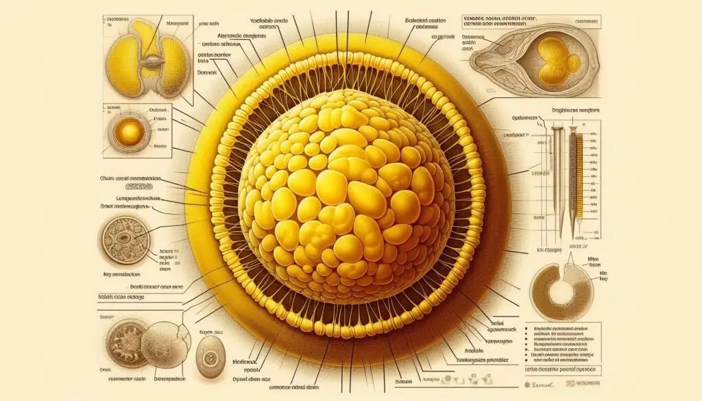
Introduction to the Yolk Sac
The yolk sac is a fascinating structure that plays a critical role during the early stages of embryonic development. It provides essential nutrients, facilitates gas exchange, and supports blood cell formation. Understanding the yolk sac’s functions helps highlight its importance in prenatal health.
What is the Yolk Sac?
It is an extra-embryonic membrane that forms during early embryogenesis. It appears shortly after fertilization and persists until about 10 to 12 weeks of gestation. During this time, it serves as a primary source of nourishment for the developing embryo. For more detailed information on embryonic structures, you can visit National Institutes of Health.
Functions of the Yolk Sac
It performs several vital functions that are crucial for the embryo’s development.
Nutritional Support
The primary role of the yolk sac is to provide nutrients to the growing embryo. It contains a rich supply of proteins and lipids that are essential for cell division and growth. Through a process called vitelline circulation, nutrients from the yolk sac are transported to the embryo via blood vessels. This process ensures that the embryo receives adequate nourishment during its earliest stages. For more information on nutrient transport in embryos, refer to ScienceDirect.
Gas Exchange
In addition to nutrient delivery, the yolk sac facilitates gas exchange. It allows oxygen to reach the developing embryo while removing carbon dioxide. This exchange occurs through its vascular system, which connects to the embryo’s circulatory system. Proper gas exchange is vital for maintaining healthy embryonic development.
Hematopoiesis: Blood Cell Formation
Another significant function of the yolk sac is hematopoiesis, or blood cell formation. The yolk sac is responsible for producing the first blood cells that are critical for embryonic development. These early blood cells help establish a circulatory system that supports growing tissues. For more insights into hematopoiesis, check out Nature Reviews.
Development of Germ Cells
It also contributes to forming germ cells, which are precursors to reproductive cells. This process is crucial for future reproductive health, ensuring that viable gametes are produced later in life.
Developmental Stages of the Yolk Sac
It undergoes several developmental phases throughout pregnancy.
Formation of the Primary Yolk Sac
The primary yolk sac forms shortly after fertilization and persists until about week 4 of gestation. It is initially large and provides significant nutritional support during this stage.
Transition to Secondary Yolk Sac
Around week 4, the primary yolk sac gives way to a secondary yolk sac. This new structure develops from mesodermal tissue and continues to support the embryo until approximately week 12. The secondary yolk sac is smaller but still plays a vital role in nutrient delivery and gas exchange.
Clinical Significance of the Yolk Sac
The size and appearance of the yolk sac can provide important indicators of pregnancy health. Healthcare providers often monitor its development through ultrasound imaging.
Indicators of Potential Complications
If it measures more than 6-7 mm or fails to appear on an ultrasound, it may indicate potential complications such as miscarriage or fetal abnormalities. Early detection can help healthcare providers take necessary actions to support maternal and fetal health. For more on ultrasound evaluations during pregnancy, visit American Pregnancy Association.
Conclusion: The Importance of the Yolk Sac
In summary, the yolk sac plays a multifaceted role in early embryogenesis by supplying nutrients, facilitating gas exchange, supporting blood cell formation, and contributing to reproductive cell development. Its functions are critical until the placenta is fully developed around week 10 to 12 of pregnancy.
More from Veterinary Anatomy:
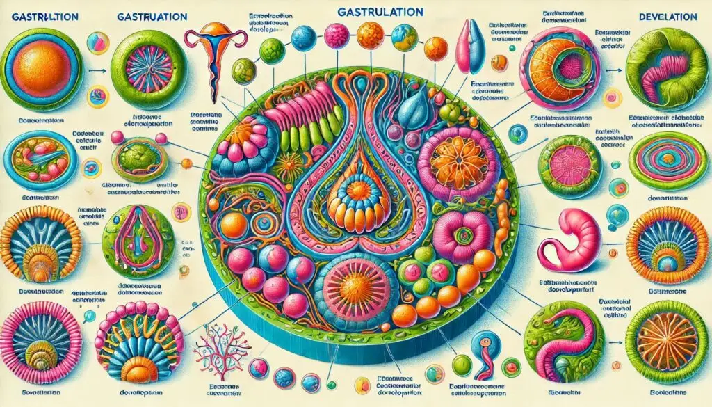
Gastrulation


ZH888game, all right! I love that I’ve found this place. Feels like a new favorite spot to unwind. Take a look at zh888game. You might like it too!