Understanding the Thoracic Cavity in Birds

Introduction
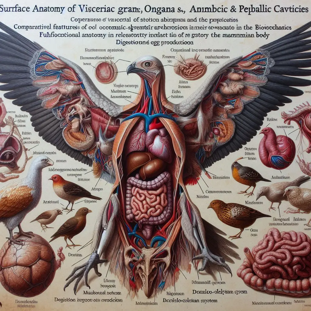
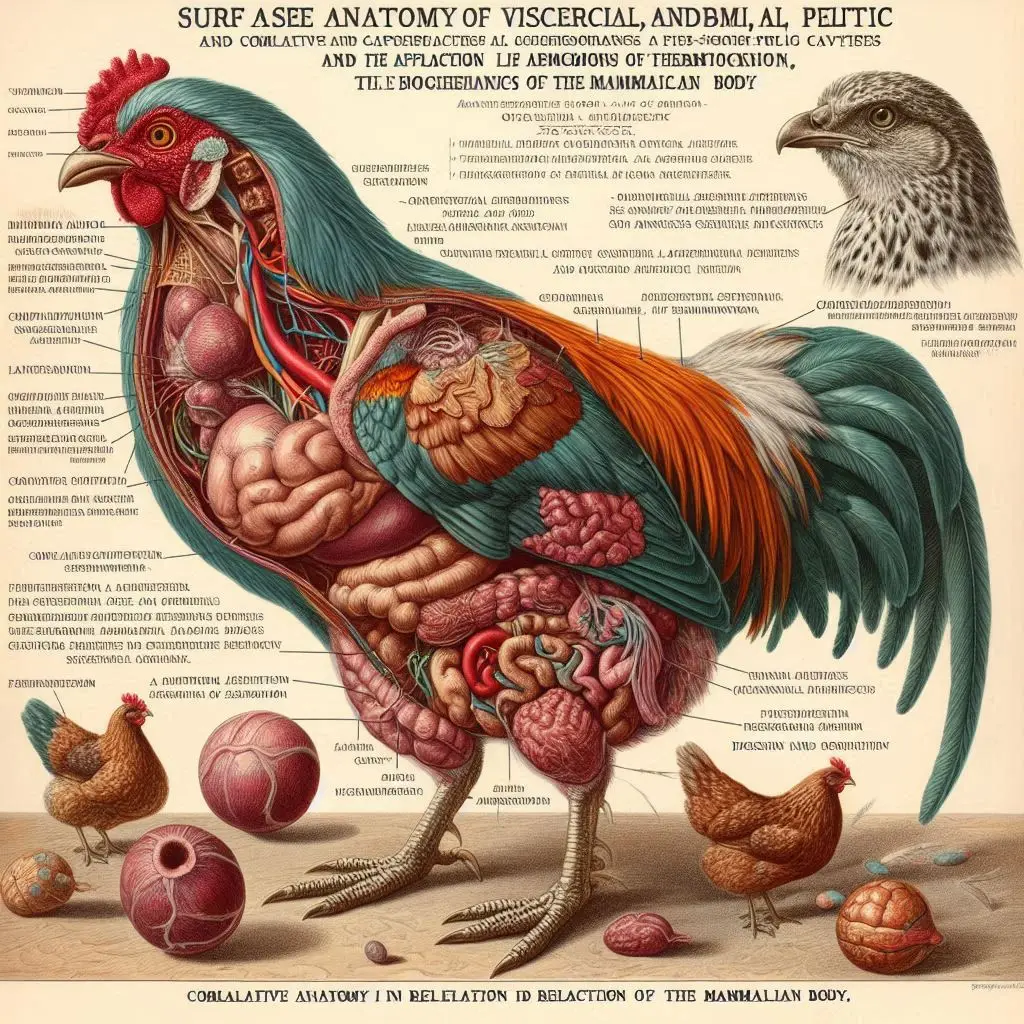
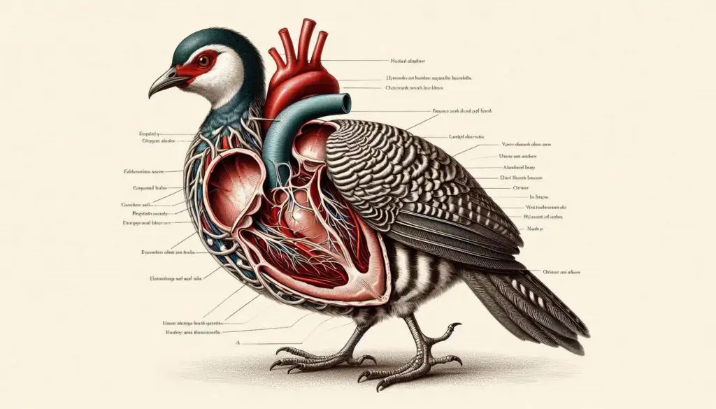
The thoracic cavity in birds is a marvel of evolution, specifically adapted for flight and respiration. This article delves into the surface anatomy of the thoracic cavity in birds, exploring its skeletal structure, muscular system, and respiratory mechanisms. Understanding these components is vital for ornithologists and veterinarians alike.
Anatomy Overview
Skeletal Structure
The skeletal structure of the thoracic cavity consists of several key components:
Sternum
The sternum, or breastbone, is a prominent feature in birds. It has a large keel that serves as an attachment point for powerful pectoral muscles. This keel is especially pronounced in flying birds, providing leverage necessary for wing movement. In contrast, flightless birds like ostriches have a flatter sternum. For more details on avian skeletal structures, check out this article from Birds of North America.
Ribs
Birds possess ribs that are unique compared to other vertebrates. Each rib has an uncinate process that overlaps with adjacent ribs. This design enhances the structural integrity of the thorax and supports the dynamic movements required during flight. The number of ribs varies among species but typically ranges from 7 to 10 pairs. You can learn more about rib structures in birds from National Geographic.
Vertebral Column
The vertebral column in birds includes cervical, thoracic, lumbar, sacral, and caudal vertebrae. The thoracic region consists of fused vertebrae that form a rigid structure known as the notarium. This fusion provides stability for the pectoral girdle during flight.
Muscular System
The muscular system plays a crucial role in avian flight.
Pectoral Muscles
The pectoral muscles are vital for wing movement and are anchored to the keel of the sternum. These muscles contract during flight strokes, allowing birds to achieve lift and maneuverability. The coracoid bone also aids in stabilizing wing position. For further reading on muscle anatomy in birds, refer to The Avian Anatomy.
Respiratory System
Birds have a highly efficient respiratory system that is essential for their high metabolic demands during flight.
Lungs and Air Sacs
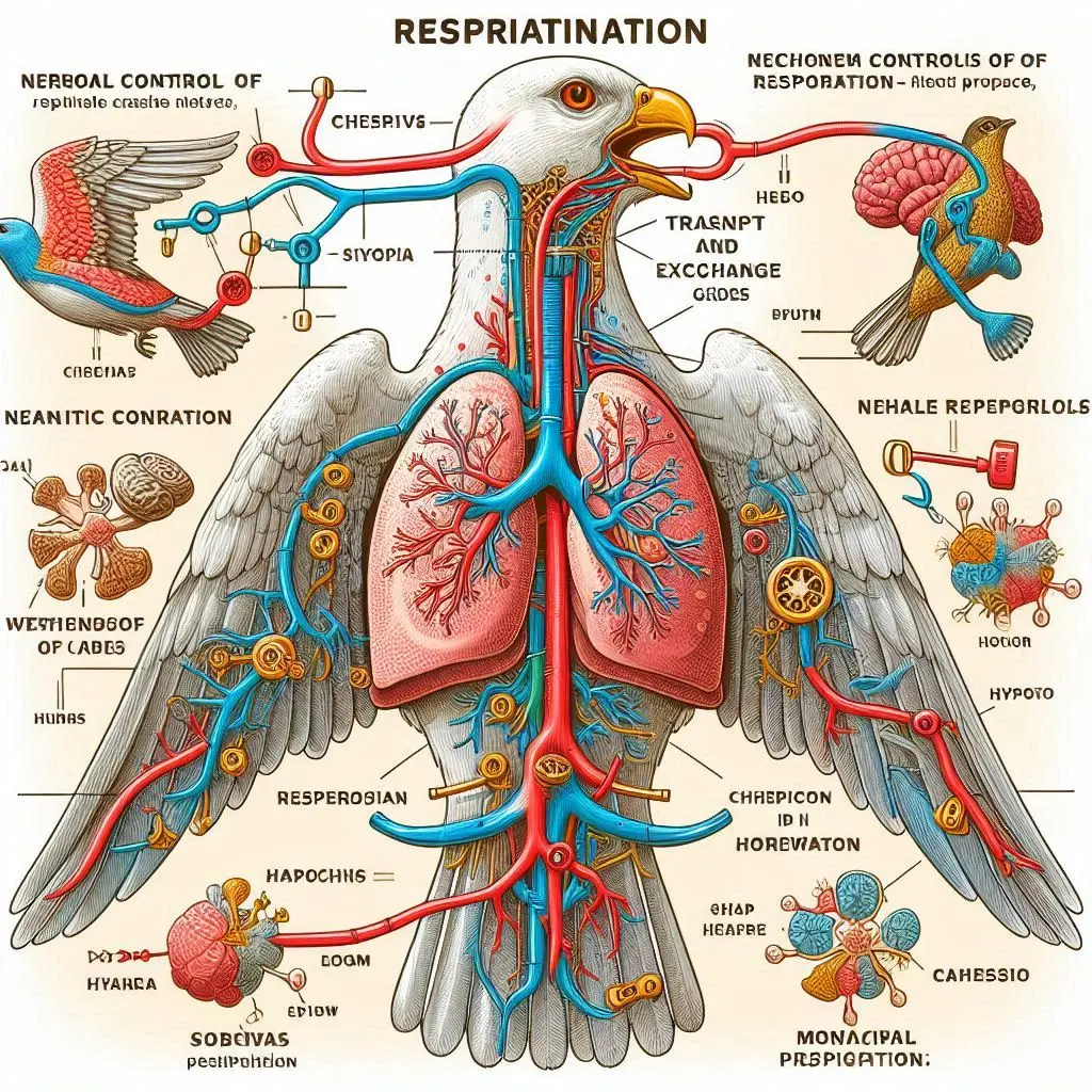
Birds possess compact lungs that are firmly attached to the thoracic wall. These lungs connect to various air sacs that facilitate unidirectional airflow. This design allows for continuous ventilation—air flows through the lungs even during exhalation.
Airflow Mechanics
The trachea bifurcates into primary bronchi leading into each lung. The secondary bronchi connect to abdominal air sacs, enabling effective gas exchange through air capillaries within the lungs. This unique system allows birds to extract oxygen more efficiently than mammals.
Functional Implications
Understanding the surface anatomy of the thoracic cavity has significant implications for various fields:
Veterinary Medicine
Veterinarians must understand avian anatomy to diagnose and treat respiratory issues effectively. Knowledge of the thoracic cavity’s structure aids in performing surgeries and managing conditions like pneumonia or air sacculitis. For insights into avian veterinary practices, visit Veterinary Partner.
Ornithology
Ornithologists study bird physiology to understand evolutionary adaptations related to flight and respiration. The design of the thoracic cavity reflects adaptations that allow birds to occupy diverse ecological niches.
Conclusion
The surface anatomy of the thoracic cavity in birds showcases remarkable adaptations for flight and respiration. From the robust skeletal structure to the efficient respiratory system, each component plays a critical role in avian biology.
More from Veterinary Anatomy:
https://wiseias.com/paraffin-embedding-tissue-processing/
https://wiseias.com/vertebrate-embryology-aves-domestic-mammals/


Responses