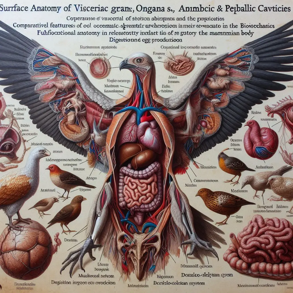
Surface Anatomy of Visceral Organs

Introduction to Visceral Anatomy
Visceral anatomy refers to the study of internal organs within the body cavities. In animals, this includes a variety of structures that perform essential functions for survival. Understanding these organs’ surface anatomy is vital for fields such as veterinary medicine, zoology, and comparative physiology.
Importance of Studying Visceral Anatomy
- Biological Insights: Knowledge of organ structure aids in understanding biological processes like digestion, respiration, and reproduction.
- Clinical Applications: In veterinary practice, recognizing normal organ anatomy helps diagnose diseases.
- Evolutionary Perspective: Studying variations across species reveals how different environments shape organ development.
The Visceral Cavity in Teleostean Fishes
Teleostean fishes, such as Eugerres mexicanus, exhibit unique features in their visceral cavities. The organization of these cavities reflects adaptations to aquatic life.
Structure of the Visceral Cavity
- Shape and Boundaries: The visceral cavity is typically oval-shaped. It is delineated by various muscular structures and bones.
- Major Components:
- Gas Bladder: This organ occupies a significant portion of the cavity and aids in buoyancy.
- Digestive Tube: Located in the lower part of the cavity alongside the liver and gonads.
- Kidneys: Positioned dorsal to the gas bladder.
- Spleen: Found among intestinal loops.
For more detailed information on teleostean fish anatomy, refer to this article on the anatomy of Eugerres mexicanus.
Functional Implications
The arrangement of organs in teleosts is closely linked to their lifestyle:
- Buoyancy Control: The gas bladder allows fish to maintain their position in water without expending energy.
- Efficient Digestion: The compact arrangement facilitates quick digestion and nutrient absorption.
Abdominal Anatomy in Large Animals
In larger mammals like cattle and camels, the abdominal cavity houses numerous vital organs. Understanding this anatomy is crucial for both practitioners and researchers.
Overview of Abdominal Structures
- Peritoneum: This serous membrane lines the abdominal cavity. It consists of two layers:
- Parietal Peritoneum: Lines the abdominal wall.
- Visceral Peritoneum: Covers most abdominal organs.
For an in-depth look at abdominal anatomy in large animals, see this resource on bovine and camelid anatomy.
Major Organs in Large Mammals
- Stomach: Ruminants have a complex stomach with multiple compartments (e.g., rumen).
- Liver: A large organ critical for metabolism.
- Kidneys: Located retroperitoneally, essential for waste filtration.
Mesenteries and Omenta
Mesenteries are folds of peritoneum that connect organs to the abdominal wall. They contain blood vessels and nerves essential for organ function. The greater omentum acts as a protective layer over the intestines.
Comparative Anatomy Across Species
The surface anatomy of visceral organs varies significantly among species due to evolutionary pressures. This section highlights key differences observed in various animal groups.
Adaptations in Aquatic vs. Terrestrial Animals
- Aquatic Animals:
- Teleosts exhibit streamlined bodies with specialized organs for buoyancy.
- Their digestive systems are adapted for processing different types of food found underwater.
- Terrestrial Animals:
- Mammals have evolved more complex digestive systems to handle a variety of diets.
- Organs are arranged to optimize space within a larger body cavity.
Evolutionary Significance
The differences in visceral anatomy reflect adaptations to environmental challenges:
- Aquatic species have developed features that enhance buoyancy and gas exchange.
- Terrestrial animals have adapted their digestive systems to maximize nutrient extraction from diverse food sources.
Clinical Relevance of Visceral Anatomy
Understanding visceral organ anatomy is essential for diagnosing diseases and performing surgical procedures in veterinary medicine.
Diagnostic Techniques
Veterinarians use various imaging techniques to assess organ health:
- Ultrasound: Non-invasive imaging used to visualize soft tissues.
- X-rays: Helpful for identifying structural abnormalities.
- CT Scans: Provide detailed cross-sectional images of internal structures.
For a comprehensive overview of abdominal viscera evaluation techniques, visit this article on anatomy of abdominal viscera.
Common Disorders Related to Visceral Organs
- Gastrointestinal Issues: Conditions like bloat or colic are common in large animals.
- Kidney Diseases: Renal failure can occur due to various factors including dehydration or toxins.
Conclusion
The surface anatomy of visceral organs plays a crucial role in understanding animal biology. From teleostean fishes to large mammals, each group exhibits unique adaptations that enhance survival. Knowledge gained from studying these structures not only aids veterinary practices but also enriches our understanding of evolutionary biology.By exploring resources like TeachMeAnatomy and StatPearls, readers can deepen their understanding of visceral organ anatomy across species.This comprehensive exploration emphasizes how vital it is to appreciate the complexity and functionality of internal organs within various animal groups.
More from Veterinary Physiology:
https://wiseias.com/abo-blood-group-system-animals/
https://wiseias.com/anticoagulation-in-animals/
https://wiseias.com/hemorrhagic-disorders-in-animals/


Responses